


Poster Presentation
Poster size is 90 cm in width and 180 cm in height.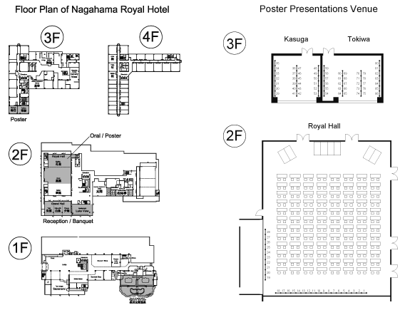
List of Poster Presentations
| P-1 | K. Ataka1, A. Baumann2, S. Kerruth1, J. Fitter2, G. Büldt2, J. Heberle1, R. Schlesinger3 (1Experimental Molecular Biophysics, Fachbereich Physik, Freie Universität Germany, 2ICS-5, Forschungszentrum Switzerland, 3Genetic Biophysics, Fachbereich Physik, Freie Universität Germany) Insertion and folding of bacteriorhodopsin into a solid-supported lipid scaffold during cell free expression: in-situ observation by surface enhanced infrared absorption spectroscopy |
| P-2 | S. P. Balashov1, L. E. Petrovskaya2, E. P. Lukashev3, E. S. Imasheva1, A. K. Dioumaev1, J. M. Wang1, S. V. Sychev2, D. A. Dolgikh2, A. B. Rubin3, M. P. Kirpichnikov2,3, J. K. Lanyi1 (1University of California, USA, 2M.M. Shemyakin - Yu.A. Ovchinnikov Institute of Bioorganic Chemistry, Russia, 3M. V. Lomonosov Moscow State University, Russia) Novel properties of the proton pump from Exiguobacterium sibiricum |
| P-3 | R. Belitzky Rozin, M. Sheves (Weizmann Institute of Science, Israel) pH-dependence of Anabaena sensory rhodopsin: retinal isomer composition, rate of dark adaptation and pKa of Asp217 side chain |
| P-4 | E. Bühl, M. Braun, A. Lakatos, C. Glaubitz, J. Wachtveitl (Goethe University Frankfurt, Germany) Wavelength dependent primary reaction of green proteorhodopsin |
| P-5 | S. K. Chan1, T. Kitajima-Ihara1, M. Murakami1, K. Ihara2, T. Kouyama1,3 (1Nagoya University, Japan, 2Nagoya University, Japan, 3RIKEN Harima Institute/Spring-8, Japan) Crystal structure of cruxrhodopsin-3 from Haloarcula vallismortis |
| P-6 | L. Chao1, G. Dai2, T. Kikukawa3, K. Ihara4, T. Iwasa1 (1Muroran Institute of Technology, Japan, 2Inner Mongolia Normal University, China, 3Hokkaido University, Japan, 4Nagoya University, Japan) Microbial rhodopsins of Halorubrum sp.ejinoor found in a salt lake in Inner Mongolia, China |
| P-7 | X.-R. Chen, C.-S. Yang (National Taiwan University, Taiwan) Investigation of the retinal-binding pocket residues affect the pH stability of acid-tolerant HmBRII from Haloarcula marismortui |
| P-8 | F.-K. Tsai1, H.-Y. Fu2, C.-S. Yang2, L.-K. Chu1 (1National Tsing Hua University, Taiwan, 2National Taiwan University, Taiwan) Photochemistry of a dual-bacteriorhodopsin system in Haloarcula marismortui: HmbRI and HmbRII |
| P-9 | K. Eisenhauer, K. Gerwert (Ruhr-University Bochum, Germany) Homology models of channelrhodopsin-2: the closed and open form of the channel |
| P-10 | T. Fujisawa1, S. Takeuchi1, S. Masuda2, T. Tahara1 (1RIKEN, Japan, 2Tokyo Institute of Technology, Japan) Signaling-state formation mechanism of BLUF proteins studied by femtosecond time-resolved absorption spectroscopy |
| P-11 | Y. Guo, V. Mooney, E. Yan (Yale University, USA) Thermal stability of vertebrate visual pigments is correlated to molecular evolution of vision |
| P-12 | N. Hamada1, N. Inazumi2, R. Nakamura1 (1Science & Technology Entrepreneurship Laboratory (e-square) and 2Department of Science, Osaka University, Japan) Influence of steric limitations around chromophore in protein cage of photoactive yellow protein |
| P-13 | T. Hasegawa1, Y. Sudo2, T. Murata3, S. Hayashi1 (1Kyoto University, Japan, 2Okayama University, Japan, 3Chiba University, Japan) Assessing molecular mechanism of high thermal stability of thermophilic rhodopsin by molecular dynamics simulation |
| P-14 | T. Hasemi1, T. Kikukawa1, M. Kamiya1, T. Aizawa1, K.-H. Jung2, N. Kamo1, M. Demura1 (1Hokkaido University, Japan, 2Sogang University, Korea) Proton transfer reactions of Anabaena sensory rhodopsin |
| P-15 | O. Hisatomi, Y. Nakatani, Y. Kai (Osaka University, Japan) A photo-activated basic-leucine zipper module, opZL |
| P-16 | M. Szczepek1, F. Beyriére1, K. P. Hofmann1, M. Elgeti1, R. Kazmin1, A. Rose1, F. J. Bartl1, D. von Stetten2, M. Heck1, M. E. Sommer1, P. W. Hildebrand1, P. Scheerer1 (1Charité - Universitätsmedizin Berlin, Germany, 2European Synchrotron Radiation Facility, France) Crystal structure of a common GPCR-binding interface for G protein and arrestin |
| P-17 | Y. Hontani1, T. Mathes1, K. Stehfest2, P. Hegemann2, J. T. M. Kennis1 (1Vrije Universiteit, the Netherlands, 2Humboldt-Universität, Germany) Investigation of photoreaction mechanism of ChR1/ChR2 with femtosecond to microsecond ultrafast spectroscopy |
| P-18 | S. Hososhima1,2,3, T. Ishizuka1,2, Y. Hiromu1,2 (1Tohoku University, Japan, 2CREST, JST, Japan, 3Research Fellow of JSPS, Japan) Bi-stable variants of chimeric channelrhodopsins ? kinetics-dependent activation of neurons |
| P-19 | K. Inoue1,2,3, F. H. M. Koua2, Y. Kato1, R. Abe-Yoshizumi1, H. Kandori1,2 (1Department of Frontier Materials, and 2OptoBioTechnology Research Center, Nagoya Institute of Technology, Japan, 3PRESTO, JST, Japan) Spectroscopic study of a light-driven chloride ion pump from marine bacteria |
| P-20 | S. Ito1, H. E. Kato2, O. Satomi3, R. Taniguchi3, T. Iwata1, O. Nureki3, H. Kandori1 (1Nagoya Institute of Technology, Japan, 2Stanford University Medical School, USA, 3The University of Tokyo, Japan) Cation effect on the photo intermediates of channelrhodopsin |
| P-21 | T. Iwata, H. Kandori (Nagoya Institute of Technology, Japan) FTIR spectroscopic analysis of the unique hydrogen-bonding formation in photoactive yellow protein |
| P-22 | S. Li, M. Jin (Louisiana State University Health Sciences Center, USA) The 26S proteasome non-ATPase regulatory subunit 13 (PSMD13) mediates rapid degradation of disease-associated mutant RPE65 proteins via the ubiquitin-proteasome pathway |
| P-23 | R. Kabutomori1, R. Sakata1, K. Okano1, Y. Kubo1, A. Takemura2, T. Miwa3, H. Yamamoto3, Toshiyuki Okano1 (1Waseda University, Japan, 2University of the Ryukyus, Japan, 3Japan Agency for Marine-Earth Science and Technology (JAMSTEC), Japan) Rhodopsin and clock genes in a snailfish, Careproctus rhodomelas, living near the deep-sea hydrothermal vent |
| P-24 | M. Kamiya, S. Hayashi (Kyoto University, Japan) A theoretical study on early intermediates of bovine rhodopsin |
| P-25 | Y. Ozaki1, T. Kawashima1, R. Abe-Yoshizumi1, H. Kandori1,2 (1Department of Frontier Materials and 2OptoBioTechnology Research Center, Nagoya Institute of technology, Japan) A color determining amino acid residue of proteorhodopsin |
| P-26 | Y. Kato1, K. Inoue1,2, R. Abe-Yoshizumi1, H. Kandori1 (1Nagoya Institute of Technology, Japan, 2PRESTO, JST, Japan) pH effect on photo-intermediate of Na+ pumping rhodopsin |
| P-27 | H. Kawaguchi, T. Nakanishi, M. Murakami, T. Kouyama (Nagoya University, Japan) Crystallographic study on the N state of pharaonis halorhodopsin |
| P-28 | A. Kawamura, Y. Yamazaki, H. Kamikubo, M. Kataoka (Nara Institute of Science and Technology, Japan) The elucidation of the relationship between β4-5 loop region and the chromophore environment in PYPs |
| P-29 | I. Kawamura1, S. Nakatani1, R. Nishikawa1, N. Kamo2, A. Naito1 (1Yokohama National University, Japan, 2Hokkaido University, Japan) Conformation and dynamics of pharaonis phoborhodopsin in the lipid environment as studied by solid-state MAS NMR |
| P-30 | R. Kazmin1, E. Ritter2, A. Rose1, M. Szczepek1, M. Elgeti3, R. Piechnick1, P. Scheerer1, P. Hildebrandt1, K.-P. Hofmann1, F. J. Bartl1 (1IMPB Charité, Germany, 2HU zu Berlin, Germany, 3UCLA, USA) Comparison of human and bovine rhodopsin activation pathways |
| P-31 | K. Shibasaki, H. Shigemura, T. Kikukawa, M. Kamiya, T. Aizawa, N. Kamo, M. Demura (Hokkaido University, Japan) The importance of Thr218 for Cl--pumping photocycle of Natronomonas pharaonis halorhodopsin |
| P-32 | N. Kimata1, M. Sheves2, R. Vogel, P. J. Reeves3, S. O. Smith1 (1Stony Brook University, USA, 2Weizmann Institute, Rehovot, Israel, 3University of Essex, UK) Hydrogen bonding changes involving extracellular loop 2 of rhodopsin suggest a mechanism for receptor activation |
| P-33 | K. Kojima1, Y. Imamoto1, R. Maeda2, T. Yamashita1, Y. Shichida1 (1Kyoto University, Japan, 2RIKEN, Japan) Rod visual pigment optimizes active states to achieve efficient G protein activation as compared to cone visual pigments |
| P-34 | E. Korchemskaya1,2, D. Stepanchikov3, A. Savchuk2 (1Institute of Physics of National Academy of Sciences, Ukraine, 2International Center “Institute of Applied Optics” of National Academy of Sciences, Ukraine; 3Zhytomir State University, Ukraine.) Holographic exploring phototransformations in E204Q bacteriorhodopsin film for constructing nanoscaled functional macromolecular blocks |
| P-35 | F. H. M. Koua1, R. A.-Yoshizumi2, H. Ono2, S. Ito2, Y. Kato1, K. Inoue1,2, H. Kandori1,2 (1OptoBioTechnology Research Center, and 2Department of Frontier Materials, Nagoya Institute of Technology, Japan) Low-temperature FT-IR/UV-vis spectroscopy and its implications in the function of the light-driven Na+-H+ pumping rhodopsin, KR2 |
| P-36 | K. Kuroi1, K. Okajima2,3, M. Ikeuchi2, S. Tokutomi3, T. Inomata4, T. Kamiyama4, M. Terazima1 (1Kyoto University, Japan, 2The University of Tokyo, Japan, 3Osaka Prefecture University, Japan, 4Kinki University, Japan) Fluctuation of TePixD plays an important role for signaling |
| P-37 | C. C. M. Lally, M. E. Sommer (Charite-Universitätsmedizin, Germany) Factors that modulate arrestin - metarhodopsin II binding stoichiometry |
| P-38 | Y. Makino1, H. Yomoda1, Y. Tomonaga1, T. Hidaka1, I. Kawamura1, T. Okitsu2, A. Wada2, Y. Sudo3, A. Naito1 (1Yokohama National University, Japan, 2Kobe Pharmaceutical University, Japan, 3Okayama University, Japan) Retinal configuration changes of sensory rhodopsin I as revealed by in situ photo-irradiation solid-state NMR |
| P-39 | H. Matsumoto1,2, T. Iwasa3, T. Yoshizawa4 (1University of Oklahoma Health Sciences Center, USA, 2Kurume University School of Medicine, Japan, 3Muroran Institute of Technology, Japan, 4Kyoto University, Japan) Non-covalent retinal β-ionone-opsin complex accelerates rhodopsin regeneration through favored proximity and orientation for Schiff base formation in solvent-inaccessible space |
| P-40 | T. Matsuyama, Y. Shichida (Kyoto University, Japan) Signaling of melanopsin, a tristable pigment |
| P-41 | M. Mehler1, A. E. Busche1, J. Kulhei1, E. Eckert2, A. Becker3, J. Becker-Baldus1, J. Wachtveitl2, V. Dötsch1, C. Glaubitz1 (1Institute of Biophysical Chemistry Goethe University Frankfurt, Germany, 2Institute of Physical & Theoretical Chemistry Goethe University Frankfurt, Germany, 3Max-Planck Institute for Biophysics, Frankfurt, Germany) Understanding helix-helix interactions in proteorhodopsin through segmental labeling and solid-state NMR |
| P-42 | J. Feng, B. Mertz (West Virginia University, USA) Validating the retinal flip of rhodopsin using molecular dynamics |
| P-43 | M. Mizuno1, A. Nakajima1, H. Kandori2, Y. Mizutani1 (1Osaka University, Japan, 2Nagoya Institute of Technology, Japan) Time-resolved resonance Raman study on structural evolution of chromophore in halorhodopsin from Natoronobacterium pharaonis |
| P-44 | T. Morizumi1, B. T. Eger1, L. Caro1, A. R. Balo1, O. P. Ernst1,2 (1Department of Biochemistry and 2Department of Molecular Genetics, University of Toronto, Canada) Observation of the binding of odorant to opsin |
| P-45 | K. Mizutani1, N. Hashimoto1, T. Tsukamoto2, Y. Sudo2,3, T. Murata1 (1Chiba University, Japan, 2Okayama University, Japan, 3JST, CREST, Japan) X-ray crystal structure of the proton pumping rhodopsin TR from the hyperthermoplile Thermus thermophilus |
| P-46 | S. Nakamura1, T. Kikukawa1, M. Kamiya1, T. Aizawa1, M. W Hahn2, N. Kamo1, M. Demura1 (1Hokkaido University, Japan, 2Innsbruck University, Austria) Photochemistry of the H+-pumping rhodopsin from freshwater Actinobacteria |
| P-47 | Y. Nakasone1, K. Okajima2, K. Hitomi3, Y. Aihara1, A. Nagatani1, J. Christie3, S. Tokutomi2, M. Terazima1 (1Kyoto University, Japan, 2Osaka Prefecture University, Japan, 3Scripps Research Institute, Japan) Time-resolved study on photoreaction dynamics of full-length phototropin from green algae |
| P-48 | R. Nishikawa1, I. Kawamura1, T. Okitsu2, A. Wada2, Y. Sudo3, N. Kamo4, A. Naito1 (1Yokohama National University, Japan, 2Kobe Pharmaceutical University, Japan, 3Okayama University, Japan, 4Hokkaido University, Japan) Functional conformation of Tyr residues in pharaonis phoborhodopsin as studied by solid-state 13C NMR |
| P-49 | Y. Nonaka1, K. Katayama1, K. Tsutsui2, H. Imai2, H. Kandori1 (1Nagoya Institute of Technology, Japan, 2Kyoto University, Japan) FTIR study of a primate blue-sensitive visual pigment |
| P-50 | K. Okano1, S. Ozawa1, H. Sato1, S. Kodachi1, T. Miyadai2, A. Takemura3, T. Okano1 (1Waseda University, Japan, 2Fukui Prefecture University, Japan, 3University of the Ryukyus, Japan) Ocular clock in the light: Photic induction and circadian oscillation of mRNAs in the fish ocular cells |
| P-51 | K. Oshima1, A. Shigeta1, Y. Makino1, I. Kawamura1, T. Okitsu2, A. Wada2, S. Tuzi3, A. Naito1 (1Yokohama National University, Japan, 2Kobe Pharmaceutical University, Japan, 3University of Hyogo, Japan) Analysis of O-intermediate trapped in the photocycle of Y185F mutant in bacteriorhodopsin by in-situ photo irradiation solid-state NMR |
| P-52 | L. Reissig1, S. Dalgleish1, T. Tsukamoto2, M. M. Matsushita1, Y. Sudo2, K. Awaga1 (1Nagoya University, Japan, 2Okayama University, Japan) Towards biological photodetectors |
| P-53 | T. Resler, B. Schultz, V. Lórenz-Fonfría, R. Schlesinger, J. Heberle (Freie Universitaet Berlin, Germany) Deuterium isotope effects on channelrhodopsin-2 |
| P-54 | M. Saita, B. Schultz, T. Resler, V. Lórenz-Fonfría, R. Schlesinger, J. Heberle (Freie Universitaet Berlin, Germany) ChR-2: The influence of the membrane and the application of a membrane potential |
| P-55 | K. Sakai, T. Yamashita, Y. Imamoto, Y. Shichida (Kyoto University, Japan) Comparative study of properties of TMT/Opn3 group opsins |
| P-56 | K. Sakurai1, A. Onishi2, H. Imai3, O. Chisaka4. T. Yamashita5, K. Nakatani1, Y. Shichida5 (1University of Tsukuba, Japan, 2Riken CDB, Japan, 3Primate Research Institute, 4Department of Biostudies , and 5Department of Biophysics, Kyoto University, Japan) Physiological analyses of mouse rods expressing chicken green cone opsin |
| P-57 | H. Tamura1, S. Chiba1, T. Furuta1, S. Sakuraba2, N. Matsubayashi2, M. Sakurai1 (1Tokyo Institute of Technology, Japan, 2Osaka University, Japan) Study of the mechanism of the light-driven ion transport in halorhodopsin based on the free energy calculations |
| P-58 | T. Kokubu, T. Yaita, T. Furuta, M. Sakurai (Tokyo Institute of Technology, Japan) Study of the photoactivation mechanisms of AsLOV2 and LOV-HTH using accelerated molecular dynamics simulations |
| P-59 | K. Sato1, T. Yamashita1, H. Ohuchi2, S. Tomonari3, A. Takeuchi4, S. Fujita-Yanagibayashi1, K. Sakai1, Y. Imamoto1, S. Noji3, A. Wada4, Y. Shichida1 (1Kyoto University, Japan, 2Okayama University, Japan, 3Tokushima University, Japan, 4Kobe Pharmaceutical University, Japan) Molecular properties of Opn5L1, a photoreceptor protein found in non-mammalian vertebrates |
| P-60 | S. Sato1, S. Miyazono2, S. Tachibanaki1, S. Kawamura1 (1Osaka University, Japan, 2Asahikawa Medical University, Japan) AL-OL coupling reaction, a possible mechanism of visual pigment regeneration in carp cones |
| P-61 | I. Schapiro1, F. Melaccio2, H. L. Luk3, M. Olivucci2,3 (1Max Planck Institute for Chemical Energy Conversion, Germany, 2Università degli Studi di Siena, Siena, Italy, 3Bowling Green State University, USA) Deciphering the photoisomerization mechanism of retinal in rhodopsin: A QM/MM Study |
| P-62 | V. Muders1, S. Kerruth2, V. Lorenz-Fonfria2, J. Heberle2, R. Schlesinger1 (1Genetic Biophysics and 2Experimental Molecular Biophysics, Freie Universität, Germany) Spectroscopic analysis of the red-shifted channelrhodopsin-1 from Chlamydomonas augustae |
| P-63 | M. Furuse1, J. Tamogami2, T. Hosaka1, T. Kikukawa3, N. Shinya1, M. Hato1, N. Ohsawa1, S. Y. Kim4, K.-H. Jung4, M. Demura3, S. Miyauchi5, N. Kamo3, K. Shimono1,5, T. Kimura-Someya1, S. Yokoyama1, M. Shirouzu1 (1RIKEN, Japan, 2Matsuyama University, Japan, 3Hokkaido University, Japan, 4Sogang University, Korea, 5Toho University, Japan) High-resolution crystal structure and photochemical properties of Acetabularia Rhodopsin I from the green alga |
| P-64 | L. Sun1, E. Kawano-Yamashita1, T. Nagata1, H. Tsukamoto2,3, Y. Furutani2,3, M. Koyanagi1,4, A. Terakita1 (1Osaka City University, Japan, 2Institute for Molecular Science, Japan, 3The Graduate University for Advanced Studies, Japan, 4PRESTO, JST, Japan) Molecular property and distribution of mammalian-like melanopsins in lamprey and hagfish |
| P-65 | Y. Suzuki1, K. Inoue1,2, R. Abe-Yoshizumi1, H. Kandori1 (1Nagoya Institute of Technology, Japan, 2PRESTO, JST, Japan) Restoration of pseudo genes of Na+ pump rhodopsin |
| P-66 | M. Takahashi1, A. Okazaki1, T. Tsukamoto2, Y. Sudo2, S. Takagi1 (1Nagoya University, Japan, 2Okayama University, Japan) Behavioral analysis of C. elegans is useful for estimating availability of novel optogenetic tools |
| P-67 | H. Tsukamoto1, D. L. Farrens2 (1Institute for Molecular Science, Japan, 2Oregon Health and Science University, USA) Energetics and conformational dynamics underlying the activation of the G protein-coupled receptor opsin assessed by site-directed fluorescence labeling and nanodisc techniques |
| P-68 | T. Tsukamoto1, Y. Sudo1,2 (1Okayama University, Japan, 2JST, CREST, Japan) Physicochemical characterization of a light-driven proton pump from an extreme thermophile |
| P-69 | S. Tsunoda1, A. Bergs1, G. Nagel2, A. Gottschalk1 (1Goethe-University, Germany, 2University of Würzburg, Germany) Optogenetic manipulation of behavior by newly engineered channelrhodopsin variants in C. elegans |
| P-70 | K. Tsutsui1, M. Otoh2, K. Sakurai2, N. Suzuki-Hashido1, T. Hayakawa2, B. J. Welker3, F. Aureli4,5, C. M. Schaffner5, L. M. Fedigan6, S. Kawamura2, Hiroo Imai1 (1Kyoto University, Japan, 2The University of Tokyo, Japan, 3State University New York Geneseo, USA, 4Liverpool John Moores University, UK, 5Universidad Veracruzana, Mexico, 6University of Calgary, Canada) Interspecific variation in ligand sensitivity of G-protein-coupled bitter taste receptors in new world monkeys |
| P-71 | S. Uyama1, T. Kitajima-Ihara1, M. Murakami1, K. Ihara2, T. Kouyama1 (1Department of Physics, Graduate School of Science and 2Center of Gene Research, Nagoya University, Japan) A chimeric proton-pumping rhodopsin designed for crystallographic analysis of light-induced structural changes |
| P-72 | A. Vogt1, P. Ranjan2, S. Kateriya2, S. Tsunoda1, P. Hegemann1 (1Humboldt-Universität zu Berlin, Germany, 2University of Delhi South Campus, India) Conversion of proton pumps into light-gated proton channels |
| P-73 | A. Wada1, T Okitsu1, N. Futamura1, Y. Niimi1, T. Ishizuka2, H. Yawo2, T. Matsuyama Hoyos3, T. Yamashita3, Y. Shichida3 (1Kobe Pharmaceutical University, Japan, 2Tohoku University, Japan, 3Kyoto University, Japan) Preparation of red-shifted PSB of retinal having enamine structure |
| P-74 | S. Wada1,2, E. Kawano-Yamashita1, S. Tamotsu2, M. Koyanagi1,3, A. Terakita1 (1Osaka City University, Japan, 2Nara Women’s University, Japan, 3PRESTO, JST, Japan) Identification of a visible light-sensitive pigment involved in the wavelength discrimination in the lamprey pineal organ |
| P-75 | H. C. Watanabe1,2, K. Welke2, M. Marazzi2, Y. Guo2, F. Beyle2, M. Elstner2, M. Sakurai1 (1Tokyo Institute of Technology, Japan, 2Karlsruhe Institute of Technology, Germany) Theoretical approach toward an understanding of molecular functions of channelrhodopsin |
| P-76 | J. Wietek1, J. S. Wiegert2, N. Adeishvili1, F. Schneider1, H. Watanabe3, S. P. Tsunoda1, A. Vogt1, M. Elstner3, T. G. Oertner2, P. Hegemann1 (1Humboldt Universität zu Berlin, Germany, 2Center for Molecular Neurobiology Hamburg, Germany, 3Karlsruhe Institute of Technology, Germany) Conversion of channelrhodopsin into a light-gated chloride channel |
| P-77 | D. Yamada1, J. Yamamoto2, I M. M. Wijaya1, T. Suzuki1, T. Iwata1, T. Ishikawa3, E. D. Getzoff4, T. Todo1, S. Iwai2, H. Kandori1 (1Nagoya Institute of Technology, Japan, 2Graduate School of Engineering Science and 3Graduate School of Medicine, Osaka University, Japan, 4The Scripps Research Institute, USA) Functional conversion of (6-4) photolyase and CPD photolyase by mutation |
| P-78 | Y. Yamazaki, Y. Mathumoto, H. Kamikubo, M. Kataoka (Nara Institute of Science and Technology, Japan) Analysis for different property of light induced structural changes between two PYPs |
| P-79 | M. Yanagawa1, M. Hiroshima1,2, T. Yamashita3, Y. Shichida3, Y. Sako1 (1RIKEN, Japan, 2RIKEN Quantitative Biology Center (QBiC), Japan, 3Kyoto University, Japan) Direct observation of higher-order oligomerization of GPCR followed by internalization |
| P-80 | P.-C. Chen, C.-S. Yang (National Taiwan University, Taiwan) Protein sequence homology of retinal-binding proteins can potentially assist biological classification in genus Haloarcula in archaea |
| P-81 | Y. Yokoyama1,2, A. Sumiyoshi3, R. Kawashima3, H. Yawo1,2, (1Graduate School of Life Sciences, Tohoku University, Japan, 2CREST, JST, Japan, 3Institute of Development, Aging and Cancer, Tohoku University, Japan) Functional MRI signal to optogenetic tactile pattern of channelrhodopsin-2 expressing rat in barrel cortex |
| P-82 | K. Yonezawa, H. Kamikubo, K. Yoshida, Y. Yamazaki, M. Kataoka (Nara Institute of Science and Technology, Japan) Structural change in Arginine 52 of photoactive yellow protein during the photoreaction |
| P-83 | K. Yoshida1, K. Inoue1,2, T. Yamashita3, R. Abe-Yoshizumi1, K. Sasaki1, Y. Shichida3, H. Kandori1 (1Nagoya Institute of Technology, Japan, 2PRESTO, JST, Japan, 3Kyoto University, Japan) Optical control of Gs-protein signalling by microbial rhodopsin chimeras |
| P-84 | K. Yoshida, H. Kamikubo, K. Yonezawa, Y. Yamazaki, M. Kataoka (Nara Institute of Science and Technology, Japan) Light-induced structural change of PYP-phytochrome related protein |
| P-85 | H. Yu1, J. Klein-Seetharaman2, S.-J. Luo1 (1Peking University, China, 2University of Warwick, UK) Genetic variation and adaptive evolution of rhodopsin in Felidae |
| P-86 | A. Zamani1,2, T. Ishizuka1,2, H.Yawo1,2 (1Tohoku University, Japan, 2CREST, JST, Japan) Light-induced membrane depolarization by two-component optogenetic |